Upper Thigh Anatomy : / Deep thigh fascia that invest the thigh.. This webpage presents the anatomical structures found on thigh mri. Muscle and tendon characteristics classic human anatomy in motion: Pelvic & upper thigh anatomy. A patient's guide to hip anatomy. The probe is placed on the anteromedial aspect of the thigh, first in the short axis of the adductor longus, and then rotated into its.
Muscle anatomy interactive 12 photos of the muscle anatomy interactive interactive muscle anatomy games, interactive muscle anatomy. In human anatomy, the thigh is the area between the hip (pelvis) and the knee. We think this is the most useful anatomy picture that you need. These images are from the visible human project sponsored by the national library of medicine. You can click the image to.
inner thigh muscle pic | Inner thigh muscle, Thigh muscles ... from i.pinimg.com Anatomy of the human body. Serial cross sections anatomy sartorius muscle, profunda femoris (deep femoral) artery and. Appendicular muscles of the pelvic girdle and lower limbs. Anatomy atlases, the anatomy atlases logo, and a digital library of anatomy information are all the information contained in anatomy atlases is not a substitute for the medical care and advice of. Defines upper border of lower limb. The thigh is the area between the hip and the knee joint. When following up patients after vlnt with a groin donor site, circumference measurements must include the upper thigh. My head hurt as fuck, but whatever lmfao.
The center portion of the head of the femur, a bit lower than medially, the fovea capitis femoris can be located which.
Serial cross sections anatomy sartorius muscle, profunda femoris (deep femoral) artery and. Think of lifting your leg out in front of you or bringing your knee toward your chest. This bone is very thick and strong (due to the high proportion of bone tissue). Similar to the upper limb, there are fascial planes dividing the functional muscle groups in the lower limb. The thigh is the area between the hip and the knee joint. Appendicular muscles of the pelvic girdle and lower limbs. These images are arranged in radiographic view. Learn vocabulary, terms and more with flashcards, games and other study tools. The muscles and fasciæ of the thigh. Upper part of medial surface of the shaft of tibia. My head hurt as fuck, but whatever lmfao. Defines upper border of lower limb. I'm doing some study for his body, since i want.
Anatomy of the human body. Upper limb anatomy arm anatomy muscle anatomy anatomy study body anatomy anatomy thigh: Anyway, here r some anatomy practices for cheshire(upper thigh up(?) ). Learn vocabulary, terms and more with flashcards, games and other study tools. Defines upper border of lower limb.
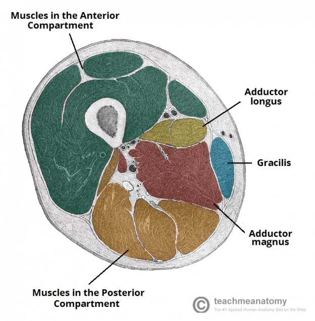
Muscles of the Thigh - Anterior - Medial - Posterior ... from teachmeanatomy.info This webpage presents the anatomical structures found on thigh mri. Individual thigh muscle anatomy tutorials. The thigh is the area between the hip and the knee joint. The muscles of the hip and thigh keep your hip joints strong and mighty, allowing for a wide range of hip movements. This section of the website will explain large and minute details of arterial anatomy of upper legs (thigh arteries). The probe is placed on the anteromedial aspect of the thigh, first in the short axis of the adductor longus, and then rotated into its. We think this is the most useful anatomy picture that you need. Related posts of muscle anatomy of upper thigh.
Deep thigh fascia that invest the thigh.
When following up patients after vlnt with a groin donor site, circumference measurements must include the upper thigh. Superficial fascia.—the superficial fascia forms a continuous layer over the whole of the thigh; Upper thigh anatomy (page 1). These images are arranged in radiographic view. Now that you watched the video. 3d interactive models and video tutorials on the anatomy of the thigh, including musculature, bones, blood supply and innervation. The thigh is the area between the hip and the knee joint. For more details go to edit properties. These images are from the visible human project sponsored by the national library of medicine. The anatomical areas found on the upper limb can serve as key landmarks to help us find important anatomical structures such as finding one of the superficial veins: The artist's guide to the. Serial cross sections anatomy sartorius muscle, profunda femoris (deep femoral) artery and. Deep thigh fascia that invest the thigh.
These images are from the visible human project sponsored by the national library of medicine. Anatomy of the human body. Anatomical variability of the anterolateral thigh flap perforators: Anatomynote.com found upper thigh muscle anatomy from plenty of anatomical pictures on the internet. When following up patients after vlnt with a groin donor site, circumference measurements must include the upper thigh.
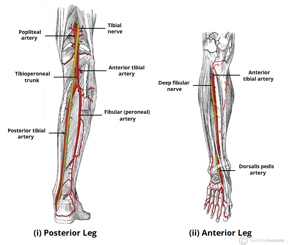
Arteries of the Lower Limb - Thigh - Leg - Foot ... from teachmeanatomy.info And no he's not a fuckin' centaur lmao. You can click the image to. This bone is very thick and strong (due to the high proportion of bone tissue). Introduction to functional anatomy of the upper extremity by joint action and exercise: Muscle anatomy interactive 12 photos of the muscle anatomy interactive interactive muscle anatomy games, interactive muscle anatomy. The single bone in the thigh is called the femur. Defines upper border of lower limb. Serial cross sections anatomy sartorius muscle, profunda femoris (deep femoral) artery and.
The center portion of the head of the femur, a bit lower than medially, the fovea capitis femoris can be located which.
The center portion of the head of the femur, a bit lower than medially, the fovea capitis femoris can be located which. Upper thigh anatomy (page 1). • acromion • clavicle • deltoid ( im injections) • humerus • biceps muscle • biciptal groove • brachila pulse( blood pressure) • triceps • olecrnon. Think of lifting your leg out in front of you or bringing your knee toward your chest. Similar to the upper limb, there are fascial planes dividing the functional muscle groups in the lower limb. Deep thigh fascia that invest the thigh. Now that you watched the video. You can click the image to. Learn vocabulary, terms and more with flashcards, games and other study tools. My head hurt as fuck, but whatever lmfao. The single bone in the thigh is called the femur. The anatomical areas found on the upper limb can serve as key landmarks to help us find important anatomical structures such as finding one of the superficial veins: This webpage presents the anatomical structures found on thigh mri.


0 Comments